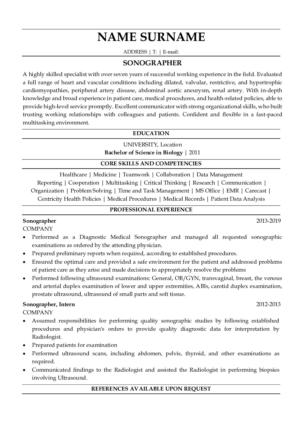
Resume Example for Sonographer
How to make a resume for a Sonographer
Ultrasonography is often a component of so-called standard diagnostic procedures. One of the most commonly performed ultrasound examinations carried out by a sonographer is an ultrasound diagnosis of the abdominal cavity. The Sonographer can do them in many cases, for example, when the patient complains of abdominal pain, weight loss, liver damage symptoms, gallstone suspicion, or kidney stones in the kidneys. When writing a resume for a sonographer in 2024, you need to describe your work experience in a similar position (Sonographer) and your professional knowledge; in particular, you can describe the progress of the study.
Job description
Ultrasonography is a method for investigating the structure of any internal organs with the help of reflected ultrasonic waves, performed by a specialist, referred to as a sonographer. This is a completely safe and painless diagnostic procedure.
An ultrasound can be performed unlimited times, including for pregnant women, without risk to health. An ultrasound study conducted by a sonographer is a more superficial method compared with magnetic resonance imaging, but often it is sufficient to diagnose various injuries. With ultrasound, the Sonographer can detect and visualize tiny and invisible other methods of studying lesion and tissue changes. Ultrasonography is a health-friendly method of studying soft tissues in the body, during which the ultrasound is used to obtain the image. All procedures are performed by the Sonographer.
For conducting all procedures, the Sonographer does not require medical attention. After the procedure, you receive photos and a conclusion from the doctor Sonographer.
Types of ultrasound examinations conducted by a sonographer:
- ultrasound of the abdominal cavity;
- ultrasound of the kidneys and urethra
- ultrasound of the prostate;
- ultrasound of the thyroid gland;
- ultrasound of the seedling;
- ultrasound of connective tissues, muscles, and joints;
- breast ultrasound.
An ultrasound study conducted by a sonographer is a non-invasive method used in prenatal diagnosis, accurately reflecting the development of pregnancy. It uses high-frequency sound waves and also allows you to get a spatial image. This gives future parents a unique opportunity to see the three-dimensional image of the future baby in the womb. During the test, the child's face is clearly visible on the screen. A performing sonographer can freely rotate the resulting image.
How does 3D ultrasound scanner work?
The device intended for 3D ultrasound scanning uses the phenomenon of propagation, scattering, and reflection of sound waves at a frequency of approximately 2-50 MHz. The ultrasound device consists of a special head - a probe (directly touching the skin in the area under study) that transmits and simultaneously receives ultrasonic waves and a computer with a monitor to which the image of the fetus is transmitted. This is possible due to the change in ultrasonic waves on an electrical impulse. Individual tissues of the human body have the ability to reflect or absorb waves to varying degrees. Electronic systems on the 3D USG device process the received data, which, in turn, leads to the creation of an image on the monitor. In addition, in comparison with traditional ultrasonic scanners, in the case of 3D research, a spatial image was obtained that allows accurate estimation of:
- anatomical data on the fetus;
- fetal bone structures.
The test course is visible on the screen in real-time and is also very readable for future parents. The doctor can stop the image at any time to measure the observed fetus. In addition, you can also print the received and save all ultrasonography on digital media.
Indications for ultrasound examination performed by a sonographer.
An ultrasound examination is carried out in order to carry out extremely detailed diagnostics of various important details. Due to high precision, ultrasound can detect a sonographer's anomaly or deformation of the fetus, which may be important in the correct therapy planning, even during pregnancy and after delivery.
What does 3D ultrasound look like?
3D ultrasound does not require special training, it is short, and its results are available immediately after completion. It is performed by a specialist sonographer lying after a preliminary detection of the abdomen. Its surface is covered with a special gel, eliminating air bubbles, which may prevent the image from being received on the monitor. The image is obtained by moving the head to the patient's skin. The test is painless.
Contraindications to 3D ultrasound
Ultrasound 3D is a completely safe diagnostic tool for the patient. There are very few contraindications for this study. Among them, you can find open wounds and large infections in the studied area.
Skills
A highly skilled, experienced sonographer provides the ability to look at the internal organs safely. The principle of image formation is limited to the transfer of pulses of ultrasonic waves. Displayed at the boundary between different acoustic environments, ultrasonic waves return with varying force and at different times to the device. The advantage of ultrasonography is real-time research that a sonographer receives in real-time on the screen, he sees an image on the camera screen that appears without a noticeable delay, and the Sonographer can appreciate the movement of structures within the body of the subject. Much valuable information can be obtained from the calculations of the ultrasound apparatus itself. You can get flow information, blood flow direction, maximum flow rate, and more detailed data.
Subscribe to our newsletter
Get $15 discount on your first order!
